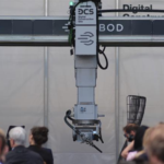
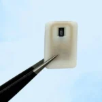
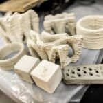
While many researchers have long been investigating a method for bioprinting a heart, this year has marked some stunning breakthroughs. Most notably scientists at Tel Aviv university were able to print a miniature heart, as we covered previously. This time around Carnegie Melon University has developed a new method they call FRESH 2, which allows them to 3D print strands of collagen. Such a method could be a massive boon to the production cardiovascular cells and collagen scaffolds.
The main issue with a lot of heart bioprinting experiments has been one of size. While the Tel Aviv researchers printed a heart, it was too small to suit a real person. Similarly, other researchers like BIOLIFE4D have mainly been doing patches and smaller scaffolds. The researchers here have successfully 3D printed five components of the human heart spanning capillary to full-organ scale. They also printed a heart valve that could open and close, and a model of a newborn heart.
“We’re nowhere near a functional heart that you can put into a human,” lead researcher, Andrew Lee stressed. “But this is an important step forward.”
Collagen Bioprinting
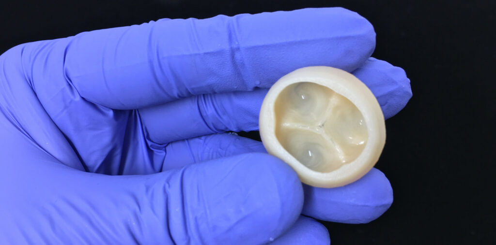
Collagen is the most abundant protein in the body. It forms a crucial part of the “extracellular matrix”, which is a network of molecules that surround your body cells, giving them structure and chemical support. This method of bioprinting avoids a major drawback in previous methods by printing living cells and soft biological material. The researchers have also stated that the approach can not only print collagen, but also other important biological substances, like fibrinogen and hyaluronic acid, so it displays a whole range of functions for bioprinting.
Other studies have found this aspect to be troublesome because collagen starts out as a fluid and deforms into a puddle when bioprinted by itself. However, within a gel, the researchers can remove collagen after the bioprinting is done. This gives it ample time to solidify. The FRESH 2 method can print tiny collagen fibers of 20 micrometers in diameter, allowing for excellent resolution.
When the researchers printed the collagen “bioink” with human heart-muscle cells, they built a small model of the heart’s left ventricle. They even displayed the ability to create micro-architectures within the body using the patient own stem cells. After a few days, the ventricles showed the capacity to contract. Such technology could yield a proper heart with further, future research.
Featured image courtesy of Carnegie Mellon University. The full study is available here.


