Researchers at UC San Diego School of Medicine and Institute of Engineering in Medicine have developed a spinal cord implant that could potentially revolutionize the way we repair nerve tissue. The implant is a 2mm replica spinal cord with a scaffolding that contains neural stem cells. Researchers recently tested it, showing improvements in regeneration in rats. With time, however, the technology could make its way to human trials, where it could aid crucial, life-changing medical procedures.
The researchers developed the implants to promote nerve growth across spinal cord injuries and improve connections. The implant focuses on axons, long, tendril-esque extensions on nerve cells that form connections to other cells. It helps organize the regenerating axons, replicating the anatomy of the pre-injury spinal cord. Experiments also used 3D printing to create scaffolds that mimic central nervous system structures. This aspect of the spinal cord implant, according to the researchers, acts like a bridge.
“In recent years and papers, we’ve progressively moved closer to the goal of abundant, long-distance regeneration of injured axons in spinal cord injury, which is fundamental to any true restoration of physical function,” said co-senior author Mark Tuszynski, MD, PhD, professor of neuroscience and director of the Translational Neuroscience Institute at UC San Diego School of Medicine.
Printing Scaffolds With Stem Cells

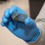
The implants are 2 millimeter in size and are the product of microscale continuous projection printing method (μCPP) tech. μCPP allowed the researchers to print 3D biomimetic hydrogel scaffolds that met the exact dimensions of the rodent spinal cord in 1.6 seconds, so the process is also lightning fast. “This shows the flexibility of our 3D printing technology,” said co-first author Wei Zhu, PhD, nanoengineering postdoctoral fellow. “We can quickly print out an implant that’s just right to match the injured site of the host spinal cord regardless of the size and shape.”
The researchers loaded them with neural stem cells and inserted them into sites of severe spinal cord injury in rats. Then, they observed that new spinal cord tissue had regrown completely where the injury was, connecting the severed ends of the host spinal cord. All of the specimen rats gained back significant functional motor improvement in their back legs. Having accomplished this feat with rats, the researchers will move the research further with larger animals eventually.
All the specimen’s circulatory systems made their way into the implants, forming functioning networks of blood vessels. This helps the neural stem cells survive. The researchers developed scaffolds with high compatibility that the host body naturally handles most of the connection repair.
Featured image courtesy of Jacob Koffler, PhD, and Wei Zhu, PhD.