
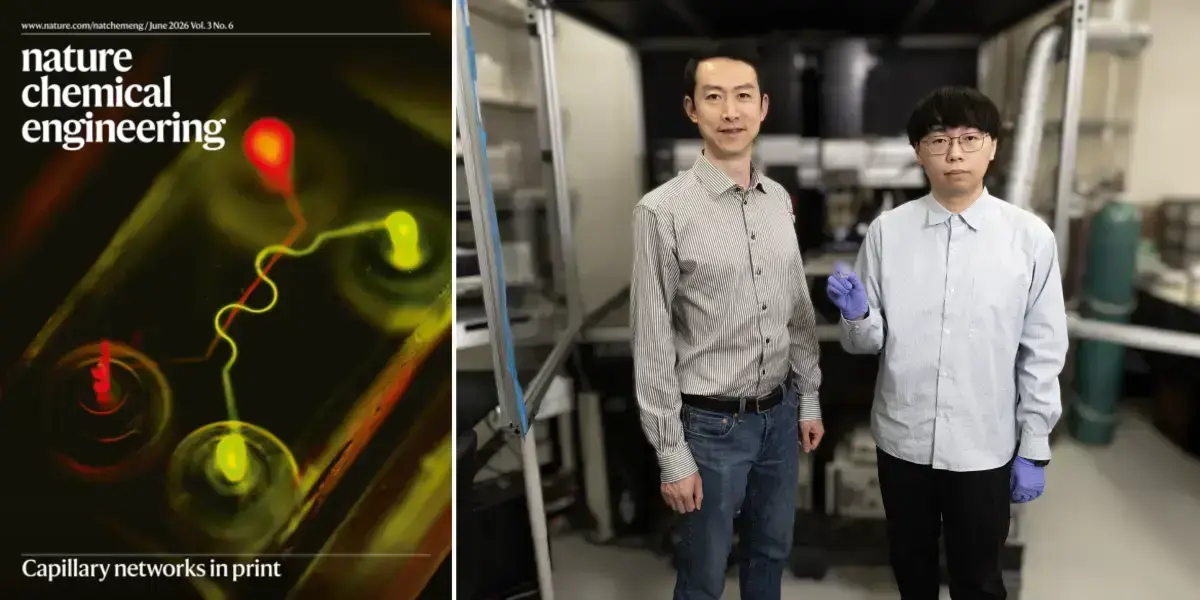
Researchers at the University of Notre Dame have developed a hybrid bioprinting technique capable of producing blood capillaries fewer than 10 micrometers in diameter, smaller than the finest human hair, clearing one of the biggest obstacles standing between scientists and lab-grown organs. The work, led by Yanliang Zhang, the Advanced Materials and Manufacturing Collegiate Professor in Notre Dame’s Department of Aerospace and Mechanical Engineering, appeared on the cover of Nature Chemical Engineering.
More than 100,000 people in the United States are currently waiting for an organ transplant, with a new candidate added every 10 minutes. Even successful transplants carry lifelong burdens: recipients must take immunosuppressive medications, face an elevated risk of infection, and live with the constant possibility of organ rejection. Bioprinting organs from a patient’s own cells would sidestep those problems, but replicating the body’s vascular networks, right down to the smallest capillaries, has stymied researchers for decades.
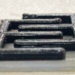
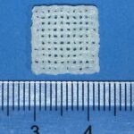
Zhang’s approach stitches together two distinct 3D printing methods. A soft, gel-like scaffold that mimics real tissue is built up layer by layer using extrusion printing. Then aerosol jet printing deposits thin gelatin threads inside the scaffold. The whole system is immersed in warm water, the gelatin liquifies and is flushed out, and the remaining channels range from hundreds of micrometers down to just a few micrometers in width.
Getting those sizes right consistently required machine learning. Small shifts in ink flow rate or sheath gas flow rate change the final channel dimensions, so the team built an autonomous optimization framework to find the right combination of parameters. “Machine learning is a very powerful tool that helps us to identify the optimal parameters much more quickly compared with the conventional trial-and-error method,” Zhang said.
Once the networks were printed, the researchers seeded select channels with endothelial cells, the same cells that line the body’s blood and lymphatic vessels. Those cells attached and spread along the inner walls, forming single-cell layers that replicate the barrier function of real human capillaries. “We are very pleased to see living cells rapidly attach and spread along the inner walls of the channels, ultimately replicating the barrier function of real human capillaries,” Zhang said. “It’s a big step forward in showing that our hybrid-printed networks are capable of supporting living tissue structures without leakage.”
The platform’s potential reaches beyond organ fabrication. Vascular models that accurately replicate capillary-scale architecture could serve as “organ-on-a-chip” test beds for evaluating drug safety and efficacy, and patient-specific cell sourcing could eventually allow therapeutic responses to be assessed in the model before full-scale treatment begins.
Zhang and collaborators at Harvard Medical School, led by Y. Shrike Zhang, associate professor of medicine at Harvard Medical School and Brigham and Women’s Hospital, have secured National Institutes of Health funding to build a more capable version of the hybrid bioprinter and push toward fabricating complete lab-grown organs, including the heart, kidney, and liver. “There are so many people waiting for an organ transplant, and the ability to print organs in the lab has the potential to transform lives by helping humans to live longer and live healthier,” Zhang said.
Source: news.nd.edu